
What is a scintillator?
A scintillator is a material—crystal, plastic, liquid, or gas—that emits light (luminescence) when struck by ionizing radiation, such as X-rays, gamma rays, or high-energy particles. These detectors absorb radiation energy, exciting electrons and releasing them as visible or ultraviolet light photons.
In the world of medical imaging and industrial inspection, X-rays are the gold standard for seeing the unseen. However, there’s a fundamental physical hurdle: CMOS sensors, the same technology you find in your smartphone camera, are incredibly good at "seeing" visible light but notoriously bad at capturing high-energy X-ray photons directly.
This is where the scintillator comes in—acting as the essential translator between high-energy radiation and digital data.
The "Translator" Mechanism
A scintillator is a specialized material (often a crystal or a phosphor layer) that absorbs X-ray radiation and re-emits that energy in the form of visible light.
When X-rays hit the scintillator, they excite its atoms. As these atoms return to their ground state, they release flashes of light (visible photons). Because CMOS sensors are optimized for the visible spectrum, they can easily "read" this light and convert it into the digital electrons that form an image.
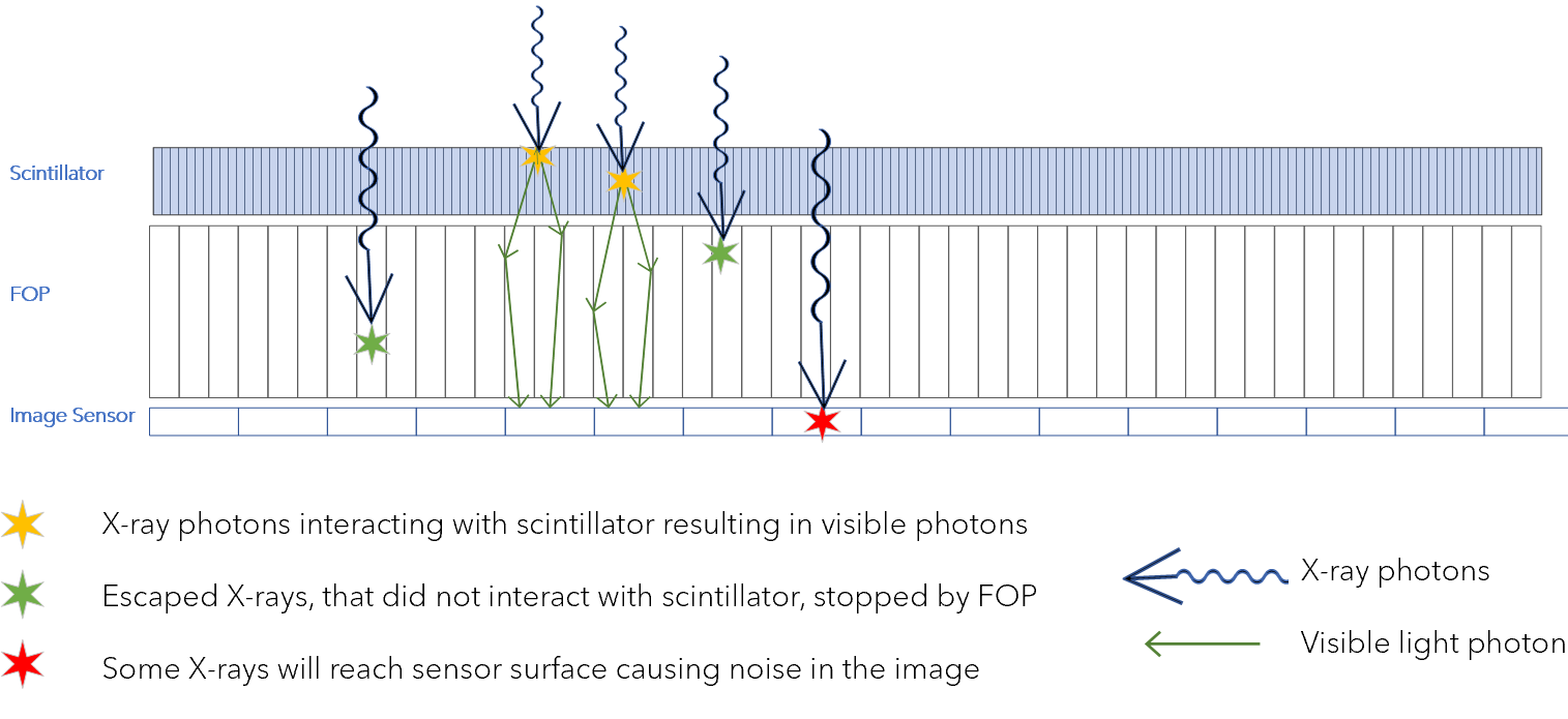
How X-rays move through a scintillator then a fiber optic plate and arrive at the image sensor in CMOS X-ray detectors
Scintillator + CMOS – a winning combination
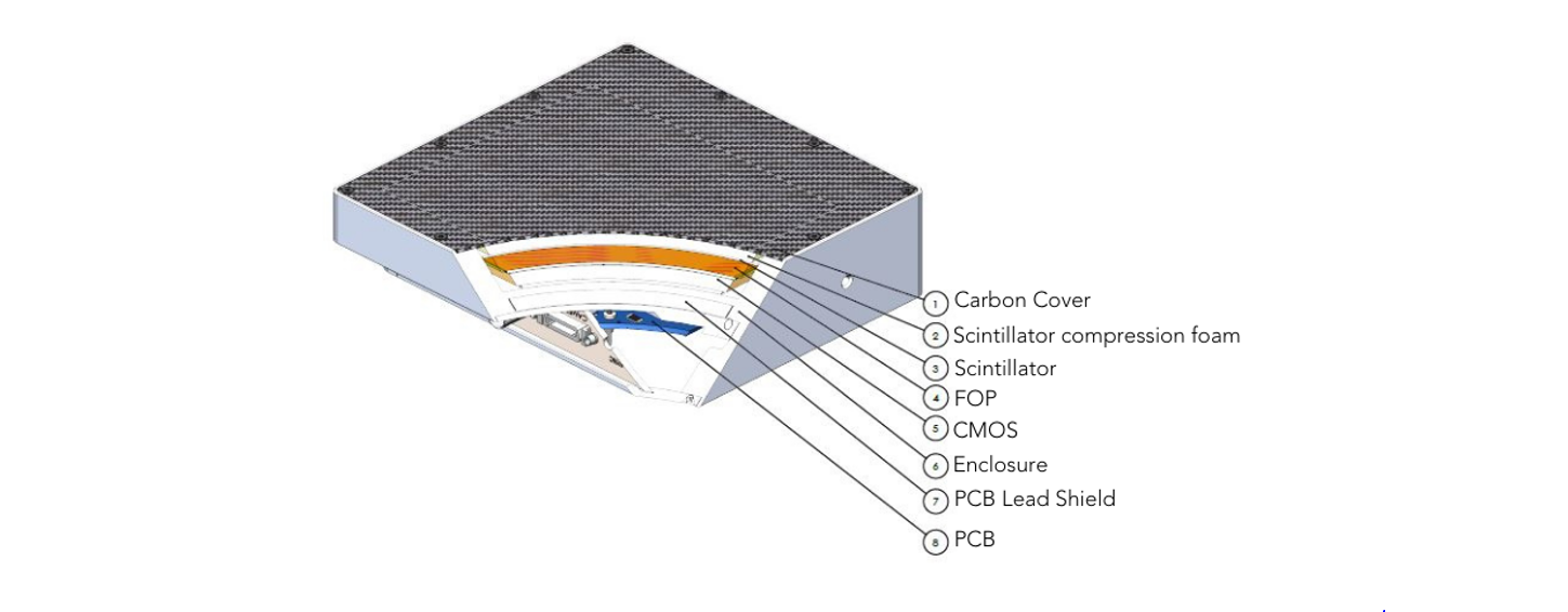
Inside a Spectrum Logic CMOS X-ray Detector
Pairing a high-quality scintillator with a CMOS sensor offers several distinct advantages over older technologies like Thin-Film Transistors (TFT) or direct conversion methods:
Ultra-High Resolution: CMOS sensors have much smaller pixel sizes than traditional aSi detectors. When paired with a "needle-structure" scintillator (like Caesium Iodide), the light is guided directly down to the pixels with minimal scattering, resulting in incredibly sharp images.
Lower Radiation Dose: Scintillators are highly efficient at stopping X-rays. Because they convert radiation so effectively, doctors can often use a lower dose of radiation to achieve a clear, diagnostic-quality image, which is a massive win for patient safety.
High-Speed Imaging: CMOS technology is fast. This combination allows for real-time "fluoroscopy" (dynamic X-rays), which is vital for guided surgeries or inspecting fast-moving parts on a production line.
Superior Signal-to-Noise Ratio (SNR): Modern scintillators are designed to match the specific visible light spectrum of CMOS sensors, ensuring that almost no information is lost in translation.
Material Matters: Caesium Iodide (CsI) vs. Gadolinium Oxysulfide (Gd2O2S)
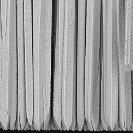
Structured Scintillator: Thallium doped micro columnar CsI
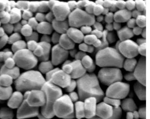
Unstructured Scintillator: Gadox
Not all scintillators are created equal. The two most common types used today are:
Caesium Iodide : Grown in microscopic needle-like micro columnar structures that channels light to prevent blurring and offer less scatter. This is the "premium" choice for high-resolution medical imaging.
Gadolinium Oxysulfide : A more cost-effective, powder type structured rugged phosphor screen. While it lacks the micro columnar structure of CsI, it is highly durable and widely used in industrial NDT (Non-Destructive Testing).
Size matters: thickness of the scintillator
A thick scintillator can be efficient against higher energy X-rays but offers less spatial resolution. A thin Scintillator can be efficient against low energy X-rays, providing better spatial resolution.
Real life AXI example – how the thickness of the scintillator can affect the energy level at which the detector can be used.
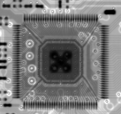
Image 1
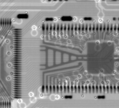
Image 2
Images acquired with 1412HR model with 600um CsI at 60kVp (High Efficiency scintillator)
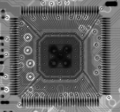
Image 3
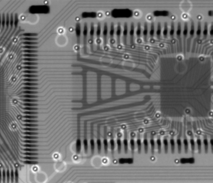
Image 4
Images acquired with 1412HR model with 290um CsI at 60kVp (High Resolution scintillator)
Images 3 and 4 (290um CsI) have better sharpness and indivudal electrical tracks and bond wires are clearly visible as compared to Images 1 and 2 (600um) CsI image.
What is MTF?
1412HR MTF High Resolution v High Efficiency Scintillators
MTF (Modulation Transfer Function).
In simple terms, MTF measures how well the detector preserves the contrast of an object as it gets smaller. Because of those micro columnar structures, CsI maintains a much higher MTF at high frequencies, which is why it’s favoured for seeing tiny fractures or fine details. Ultimately the choice of scintillator often comes down to the MTF and efficiency.
Conclusion
Without the correct scintillator, the high-speed, high-resolution capabilities of CMOS sensors would be wasted on X-rays. By converting the invisible into the visible, this duo provides the clarity and speed required for the next generation of dental, medical, and industrial diagnostics.
Scintillator Comparison: CsI v Gd2O2S
| Feature | Caesium Iodide (CsI:TI) | Gadolinium Oxysulfide (Gd2O2S) |
|---|---|---|
| Structure | Columnar (Needle-like) – acts like fiber optics to guide light. | Turbid (Granular) – light scatters more easily. |
| Spatial Resolution | High – minimal light spread means sharper edges. | Moderate – scattering can cause slight blurring. |
| Light Output | High – very efficient at converting X-rays to light. | Lower – requires slightly more radiation for same signal. |
| Durability | Delicate – sensitive to moisture (hygroscopic) and shock. | Robust – very stable and resistant to environmental factors. |
| Cost | Higher – complex manufacturing process. | Lower – more economical for large-scale detectors. |
| Primary Use | Medical imaging, Dental, Mammography. | Industrial NDT, Security (Luggage) scanning, Veterinary. |
For more information about our products and which scintillator is best for your application please contact us on inquiries@spectrumlogic.com.
Images of scintillators courtesy of Scintacor